
How the Human Eye Works
Seeing is possible thanks to a complicated series of events that start in the eyes and end in the brain. The entire process happens almost instantaneously and is only successful if every part of your visual system works properly.
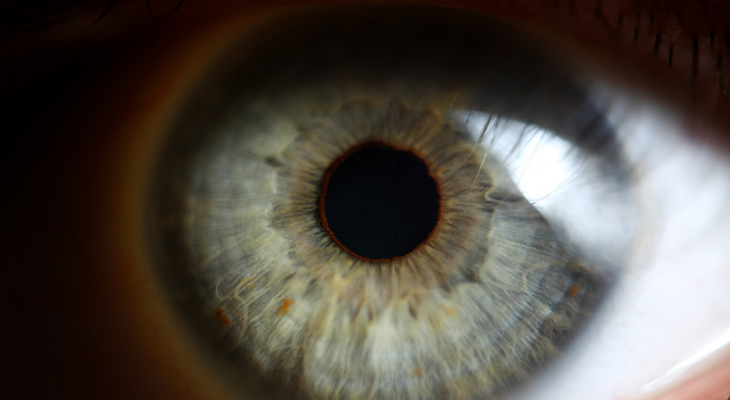
It All Starts with the Cornea
Your cornea, a clear, rounded layer of tissue that covers your pupil and iris, helps light reach your eyes by bending the rays as they enter your pupils.
The iris and pupil work together to let light into the eye. Have you noticed that your pupils look bigger when the light is dim? Tiny muscles in the iris make the pupil bigger when it's harder to see, allowing more light to enter your eyes. The muscles shrink the size of your pupils when it's bright outside or inside.
The Lens Is Essential for a Clear Picture
Light rays pass through the lens and the vitreous humor after entering the eye. The lens is a transparent disc located inside the eye under the iris and pupil, while the vitreous is the clear gel that gives the eye its shape.
The muscles that control the shape of the lens relax, causing the lens to flatten when you look at an object in the distance. The opposite happens when you look at something nearby. The muscles contract, thickening the lens. The ability of the lens to change shape allows you to shift your focus from near to far objects and back again.
The lens focuses light rays on the retina in the back of the eye. If your eyeball is too long, the rays will focus in front of the retina, causing myopia, or nearsightedness. If you're nearsighted, close objects are easy to see, while everything in the distance looks blurry.
Hyperopia, or farsightedness, occurs when your eyeball is too short. Light rays focus beyond your retina and make your near vision blurry.
As you get older, the lenses of your eyes become less flexible. The loss of flexibility affects your ability to see near objects clearly. This condition, called presbyopia, usually begins in your early- to mid-forties, according to the American Optometric Association. Fortunately, a pair of reading glasses will make it much easier to read a book or thread a needle.
The Retina Transforms Light Rays
The retina covers the back of the eye and contains two types of photoreceptor cells. The rods, found in the outer part of the retina, are essential for side vision and help you see in dim light. Cones, the other type of photoreceptor cells, are located in the macula, the center part of the retina. Cones are needed for color vision and also help you see fine details easily.
The retina turns light rays into electrical impulses, then sends the impulses to the occipital lobe of the brain through the optic nerve. Problems with your retinas or optic nerve can affect your vision.
If you have macular degeneration, a common age-related eye condition, your central vision may be blurry or you may notice blank spots in the middle part of your vision. Increased eye pressure due to glaucoma can damage your optic nerve, causing partial or total loss of vision.
How Your Brain Helps You See
The visual cortex in the brain's occipital lobe serves as the vision command center. The cortex processes and stores images and helps you make sense of what you see. Thanks to your brain, you can recognize faces, colors, letters, and shapes. You can also see fine details, avoid obstacles, keep your balance, predict the speed of moving objects, remember the things you've seen in the past, and easily recall information after reading it.
Your brain also controls the muscles that move your eyes and turns the slightly different information received from each of your eyes into one clear image.
If a stroke, tumor, infection, disease, or head trauma injures the brain, your vision and your ability to understand what you see can be temporarily or permanently affected.
Annual visits to the optometrist help you ensure that every part of your visual system is working properly. Contact our office if you're ready to schedule your next eye exam.
Sources:
American Optometric Association: Adult Vision: 41 - 60 Years of Age
National Eye Institute: How the Eyes Work
American Optometric Association: How Your Eyes Work
American Macular Degeneration Foundation: How the Eye Works as a Camera
American Academy of Ophthalmology: Eye Anatomy: Parts of the Eye and How We See, 3/9/21