Every type of doctor’s visit has a “How about not today?” part. At the eye doctor, it is dilation drops. We get it. When instilled on the eyes, the drops have a burn to them (although I think Minnesota mosquitos bite harder). For several hours after the exam, vision can be a little off (especially reading for those under 50), and it is uncomfortable to be in well-lit spaces or outside without sunglasses. I am not going to force you to be dilated; it is your day and your choice. But before you vow to never let your eye doctor dilate your eyes ever again, hear me out first.
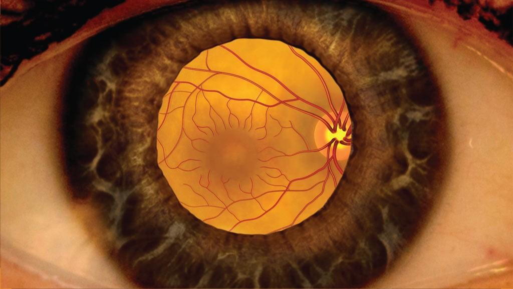
There are two parts to your eye exam. Vision (“Which is better 1 or 2?”) and eye health (think bright lights that you swear must be bad for you). Examining the health of the front of your eyes is easily accessible. What limits the ability to look inside the eye is the size of your pupil, the black opening that gets smaller with increased light, but the inside of the eye is where vision happens. It’s there that the retina captures light, allowing you to see a car pull into the lane next to you. It’s where your eye, with or without glasses or contact lenses, perfectly focuses light onto the macula, allowing you to see the faces of your loved ones clearly. It’s where your optic nerve transmits the images of the faces of your loved ones and the car next to you back to the brain. This is where blood vessels fuel the retina to make vision possible.
Without dilation drops, I only see a fraction of the inside of your eye. It’s like trying to find a key in a dark room by looking through a keyhole in the door with a flash light. You might miss the key! Wouldn’t it be easier to open the door and walk around with the flashlight? That is what dilation drops allow us to do!
How often should you have your eyes dilated? It depends on probability. What are your chances of having a vision-threatening or life-threatening issue that needs to be discovered? In young healthy patients, that risk is low. A child should have their eyes examined at 6 months, 3 years, and once before school. At one of those early eye exams, dilation should occur to look for rare congenital cancers and/or if we suspect they have an abnormally high prescription or an eye turn that could disrupt the vision development in the brain. After that? Every two to four years may be ok for most. Pupils are large when we are young, making it easier to examine most of the important structures in the eyes without drops. As we get older, pupils get smaller. Over 60 years of age, the recommendations are to dilate annually as vision-altering diseases like cataracts, glaucoma, macular degeneration, and retinal detachment are more likely. If you have a medical issue that can affect the blood vessels in your body, like diabetes or high blood pressure, we should dilate annually to evaluate if retinal blood vessels are being damaged.
We also offer retinal photographs to help us look for vision-threatening diseases on the day of your eye exam and to monitor for subtle changes that can be caught earlier by comparing it to past photographs. If you can’t be dilated that day, these photographs offer an exceptional view of the retina. Photographs don’t replace dilation, however. Even the best cameras don’t see everything. The best way to ensure an important finding is not missed is a dilated eye exam and a photograph. Photographs turn the light on in the dark room!
One patient of mine comes to mind every time someone declines to be dilated. I often talk about her when I am on the road teaching a board preparation course to optometry students or continuing education to other optometrists to drive this point home. This 52-year-old female had no issues but did have diabetes and high blood pressure - it was high that day too. She preferred not to be dilated despite educating her on how diabetes and high blood pressure can harm her retina. It had been several years since she had it done. When looking through her small undiluted pupil (key hole), I happened to view a retinal hemorrhage. Then another one. Then another. I ordered a photograph of the retina to get a better view. She had a hemi-retinal vein occlusion likely secondary to her high blood pressure; half of her retina was unable to drain its blood. This condition can cause blindness if not caught early and monitored! There are countless other examples of vision-threatening findings caught due to being dilated and/or having a photograph present.
We don’t want to disrupt your day, but we want you to see your best AND maintain the vision you have. Sometimes that means wearing our not so cool dilation sunglasses for a few hours. PS: bring your own cool sunglasses to every eye exam!
By: Dr. Jordan Keith