

Diagnostic Imaging Tests We Perform

The Ophthalmic Imaging Center at RAWNY uses state of the art equipment and techniques to image the eye for documentation and diagnostic purposes. These imaging tests include high-magnification and widefield fluorescein angiography, ICG angiography, stereo fundus photography, fundus autofluorescence, high definition optical coherence tomography, and ultrasonography.
Results are promptly available and interpretative reports by our physicians are also available as needed. As a leading provider of ophthalmic imaging in the community, our service accepts referrals from numerous ophthalmologists in Rochester and its surrounding areas.
The staff at the Ophthalmic Imaging Center are highly skilled specialists who are committed to producing the highest quality imaging. They hold clinical trial imaging certifications from the University of Wisconsin Fundus Photography Reading Center, the OCT Reading Center at Duke, the Digital Angiography Reading Center of New York, Doheny Institute Reading Center, Boston Image Reading Center, and the Optic Nerve Research Center.
Fluorescein Angiography
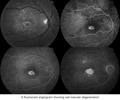
A fluorescein angiogram is a diagnostic test that helps your ophthalmologist see what is happening in your retina, highlighting any abnormalities that may be present. It is used most often to monitor two common conditions: age-related macular degeneration (AMD) and the effects of diabetes on the eyes, but can be used to detect other retinal problems as well. The images from fluorescein angiography help your doctor decide on the best course of treatment for your condition.
During a fluorescein angiogram, a harmless fluorescent dye is injected into a vein in your hand or arm, where it travels throughout the blood vessels in your body, illuminating them. As the dye passes through the blood vessels in the eye, a special, high-magnification camera takes photographs of the retina. This procedure takes about 15 minutes and is done in the office.
Side effects include:
- Your eyes may be sensitive to light due to the dilating eye drops. Sunglasses may be helpful.
- Your vision may be blurry due to the dilating eye drops. This may last a few hours.
- Your vision may appear darker or have a color tint for a few minutes afterwards.
- After the fluorescein dye is injected, your skin may turn yellowish for several hours.
- Because your kidneys remove the dye from the body, your urine will turn dark orange or yellow for up to 24 to 48 hours following the test.
Allergic reactions to fluorescein dye are rare. If they do occur, they may cause a skin rash, itchy skin, or breathing difficulty. This is usually treated with oral or injectable antihistamines.
Fundus Photography
During your visit, your physician may ask you to have some fundus photographs taken of your eye. "Fundus" refers to the back, inner lining of the eye, or the retina. Fundus photography is a highly specialized form of medical imaging that is used to document the condition of the retina, macula, blood vessels, and the optic nerve. The pictures are used for documentation, comparison, and also used to diagnose certain eye conditions. For the patient, this process is easy and relatively quick.
A special kind of camera, called a fundus camera, is used for these photographs. This camera is essentially a microscope turned on its side, with a digital camera attached to it. The photographer uses high-powered lenses to focus on the retina through the cornea, pupil and lens.
The eyes must be fully dilated in order to get good pictures. This allows for maximum amount of light to penetrate the retina, and also so the pupils do not constrict after the bright flashes of light. The patient is asked to stare at a small fixation light. The patient will then see a series of flashes of light, as the photographer takes the pictures. These photographs typically take 5 to 10 minutes.
Optical Coherence Tomography (OCT)
Optical Coherence Tomography (OCT) is a non-invasive, diagnostic test commonly used to image the retina, specifically the macula and optic nerve. Tomography refers to creating an image of a sectional plane within a body. OCT is a technique for high-resolution, cross-sectional imaging that allows your physician to diagnose, monitor and document retinal disease.
OCT provides extensive retinal analysis along with an age-matched normative database for comparison. The retinal nerve fiber layer (RNFL) and macular thickness can be precisely measured on a microscopic level. Measurement of the optic nerve head and RNFL can detect and monitor glaucoma. Furthermore, OCT can be used to image macular holes, macular edema, epiretinal membranes, macular degeneration, and other retinal pathology. OCT imaging is especially helpful when comparing pre and post-operative conditions of the retina.
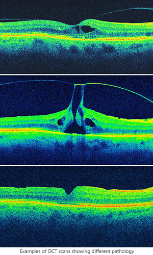
OCT imaging can be likened to an ultrasound, except light waves are used instead of sound waves. The light waves are reflected off of the retinal layers to create a detailed, cross-sectional image. Different densities in the retinal layers are identified by different colors on the scan, creating a colorful representation of the retinal layers along with any abnormal pathology. The newest OCT technology images the retina at a near cellular level, providing more detail and information than ever before. RAWNY is pleased to offer spectral domain, high-definition OCT.
Ultrasonography
Ultrasonography, also called a B-scan, is an imaging technique that is used to visualize the retina or other intraocular structures, through media opacities that make a clinical exam difficult or impossible. For example, a dense hemorrhage filling the eye may obstruct the view of the retina, prohibiting the view of a possible detachment or tear.
Situations where a direct view of the retina may be difficult include dense cataracts, eye lid problems, corneal opacities or scars, and vitreous debris or hemorrhage. The physician will use an ultrasound to look for and document detachments, tears, vitreous hemorrhages, severe edema, and also for measuring intraocular tumors.
Ultrasonography uses high-frequency sound waves, which are transmitted from a probe into the eye. As the sound waves strike intraocular structures, they are reflected back to the probe and converted into an electric signal. The signal is subsequently reconstructed as an image on a monitor, which can be used to make a dynamic evaluation of the eye or can be photographed to document pathology.
During an ultrasound, a small probe is placed on the patients eyelid or directly on the eye. The probe is moved around to image the area of interest. This diagnostic test is painless and usually takes 5 to 10 minutes.